Rhabdomyolysis: Causes, Symptoms, Lab Tests and Treatment
Reviewed by the LabReadAI medical team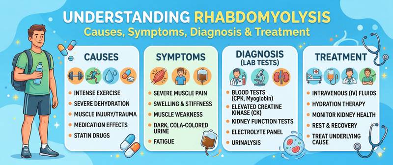
Dark tea- or cola-coloured urine after an intense workout, trauma, or prolonged immobility is one of the most recognisable warning signs of rhabdomyolysis. Behind this symptom lies the mass destruction of muscle cells, the release of toxic myoglobin into the bloodstream, and a real threat of acute kidney failure. Rhabdomyolysis can be mild and pass unnoticed — or fulminant, with fatal outcome without emergency intervention.
What Rhabdomyolysis Is and Why It Leads to Kidney Failure
Rhabdomyolysis is the breakdown of skeletal (striated) muscle tissue with the release of intracellular contents into the systemic circulation. Muscle cells (myocytes) lose membrane integrity — and myoglobin, potassium, phosphorus, uric acid, intracellular enzymes (primarily creatine kinase, CK), and other substances enter the blood.
The primary danger is myoglobin. In the renal tubules, myoglobin precipitates in acidic urine, forms casts, and causes direct tubular toxicity. The result is acute kidney injury, developing in 15–50% of patients with significant rhabdomyolysis. In addition, the massive release of potassium from necrotic myocytes threatens life-threatening hyperkalaemia and cardiac arrest.
Causes of Rhabdomyolysis: From Exercise and Statins to Trauma
Rhabdomyolysis can develop in vastly different circumstances — from the gym to the intensive care unit.
Traumatic and physical causes:
- Crush syndrome — prolonged muscle compression (earthquakes, road accidents, prolonged unconscious lying)
- Excessive physical exertion — "exertional rhabdomyolysis": marathons, intense CrossFit, military training. Especially dangerous in heat and dehydration
- Seizures — prolonged epileptic status
- Hyperthermia and heat stroke — overheating causes direct thermal myocyte injury
- Electrical trauma and lightning strike
- Muscle ischaemia — arterial thrombosis, positional compression syndrome
Non-traumatic causes:
- Drugs and toxins — the most common non-traumatic cause:
- Statins — in 0.1–0.5% of patients; risk rises sharply when combined with fibrates, ciclosporin, amiodarone, or CYP3A4 inhibitors (clarithromycin, itraconazole)
- Alcohol — direct myotoxicity and hypokalaemia
- Cocaine, amphetamines, MDMA (ecstasy)
- Toxins — snake and spider bites
- Infections — viral myositis (influenza, HIV, enteroviruses), bacterial sepsis
- Metabolic disturbances — hypokalaemia, hypophosphataemia, hyponatraemia, hypothyroidism
- Genetic myopathies — carnitine palmitoyltransferase II deficiency, glycogen storage diseases, malignant hyperthermia
- Autoimmune myositis — polymyositis, dermatomyositis
Rhabdomyolysis Symptoms After Exercise: The Classic Triad
The classic clinical picture of rhabdomyolysis consists of three features in combination:
1. Muscle pain and weakness — most often in muscles subjected to injury or exertion. Pain may be localised or diffuse, from mild to severe. Muscles may be swollen and tender on palpation.
2. Dark urine — brown, red-brown, or "tea-coloured" / "cola-coloured" — the hallmark of myoglobinuria. It appears when myoglobin concentration in the urine is high enough to visibly change colour. Important: in some patients, urine colour does not change — rhabdomyolysis without visible myoglobinuria occurs.
3. General symptoms — malaise, fever, tachycardia, nausea, reduced urine output (oliguria) when AKI develops.
Danger signs requiring immediate hospitalisation:
- Anuria or critically reduced urine output
- Cardiac arrhythmia (hyperkalaemia)
- Progressive weakness or confusion
- Limb swelling with pain and sensory loss — compartment syndrome
Blood Tests for Rhabdomyolysis: CK, Myoglobin and Key Markers
Blood:
CK (creatine kinase) — the primary rhabdomyolysis marker. Normal < 200 U/L. In rhabdomyolysis, CK rises tens to hundreds of times: levels of 10,000–100,000 U/L are typical; in severe cases > 100,000 U/L. CK peaks 24–72 hours after the onset of injury and then declines at approximately 50% per day once injury stops.
Creatinine — rises with developing AKI. Key nuance: in rhabdomyolysis, urea rises faster than creatinine (accelerated protein catabolism), and the urea/creatinine ratio may be higher than usual.
Potassium — hyperkalaemia from massive K⁺ efflux from destroyed cells. Life-threatening levels (> 6.5 mmol/L) require emergency treatment.
Phosphorus — hyperphosphataemia from phosphate release out of myocytes.
Calcium — hypocalcaemia in the acute phase (calcium binds to damaged muscle tissue); paradoxical hypercalcaemia in the recovery phase (calcium returns from muscle to blood).
Uric acid — elevated due to purine release from destroyed cells.
AST — elevated (AST is present not only in the liver but also in muscle; in rhabdomyolysis — muscle-source elevation). ALT rises much less — a sharp AST rise with near-normal ALT points to muscle rather than liver origin.
LDH — markedly elevated: like CK, it is an intracellular enzyme released in large quantities when myocytes die. In rhabdomyolysis, LDH rises in parallel with CK and serves as an additional marker of the severity of muscle tissue damage.
Urine:
- Urine dipstick positive for blood (reacts to myoglobin the same as haemoglobin), but red blood cells are absent on microscopy — the classic discrepancy pointing to myoglobinuria
- Brown sediment, granular casts
- Urine myoglobin (direct assay) — confirms the diagnosis, but a negative result does not exclude it
Treatment of Rhabdomyolysis
Aggressive intravenous fluid therapy — the cornerstone of treatment. Goal: prevent AKI by "flushing" myoglobin from the tubules and maintaining high urine output.
- Volume: 1–1.5 L/hour in the first hours — in hospital under urine output and haemodynamic monitoring. Target urine output: 200–300 mL/hour until CK normalises
- Solution: isotonic saline (0.9% NaCl) — first line. Adding sodium bicarbonate (urinary alkalinisation) theoretically reduces myoglobin precipitation, but clinical superiority over saline alone is not established
- Mannitol — discussed as an osmotic diuretic but is not a standard recommendation
Eliminate the cause:
- Discontinue the offending drug (statin or other)
- Treat the underlying condition (infection, seizures)
- In crush syndrome — surgical decompression for compartment syndrome
Manage complications:
- Hyperkalaemia — standard protocols (calcium gluconate, insulin + glucose, dialysis)
- AKI — renal replacement therapy when refractory
- Compartment syndrome — fasciotomy
Prognosis: with mild rhabdomyolysis and early aggressive hydration — full recovery. With severe rhabdomyolysis and AKI — ICU mortality 3–5%; significantly higher with multiorgan failure.
When to Seek Urgent Medical Attention
- Dark urine after intense exercise, trauma, electric shock, or prolonged lying — go to the emergency department immediately
- Severe muscle pain and weakness after exertion in the heat — do not wait if urine darkens
- Reduced or absent urine output — sign of developing AKI
- Arrhythmia, weakness, or numbness — possible hyperkalaemia; call emergency services
- Starting statins + unexplained muscle pain and weakness — check CK and see a doctor
This article is for informational purposes only and does not replace consultation with a qualified physician.
For informational purposes only
This article is for informational purposes only and does not constitute medical advice, diagnosis, or treatment. Please consult a healthcare professional for medical guidance.