Chest Pain: Causes, Diagnosis and When to Call Emergency
Reviewed by the LabReadAI medical team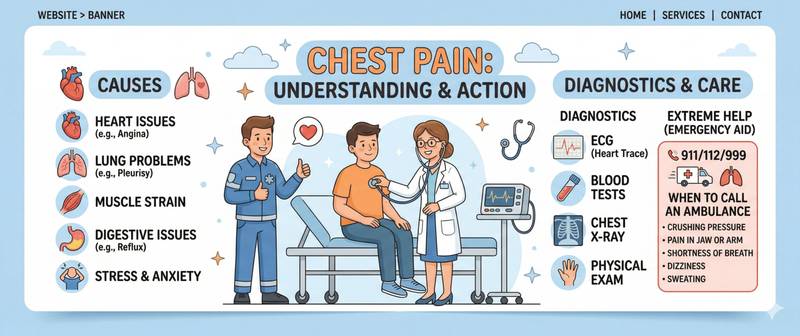
Chest pain is one of the most frightening symptoms — and one of the most ambiguous. Behind it can lie a life-threatening heart attack or a simple intercostal muscle spasm. Reading the situation correctly within the first minutes can save a life or prevent unnecessary panic. This article covers the causes, diagnostic tests, and the central question: when to call emergency services.
Why the Chest Hurts: The Anatomy of the Symptom
The chest is a complex, multi-layered structure. Pain here can originate from dozens of sources: the heart, lungs, pleura, oesophagus, stomach, ribs, cartilage, intercostal muscles, nerve roots, and even the skin. The brain does not always accurately "address" the pain signal: cardiac pain frequently radiates to the shoulder, jaw, or arm — a phenomenon called referred pain. Pain from the oesophagus can mimic angina in extraordinary detail.
This is precisely why self-diagnosis of chest pain is dangerous in both directions. Someone having a heart attack may convince themselves it is "the stomach" or "a pulled muscle" — and loses critically important time. Someone with intercostal neuralgia may wait for an ambulance they do not need.
The key principle: with any chest pain of unknown cause — rule out the life-threatening first, then look for benign causes. Never the other way around.
High-risk individuals — patients with atherosclerosis, those with diabetes, smokers, people with obesity and hypertension, elderly men — should have chest pain evaluated by a cardiologist as the default.
Cardiac Causes of Chest Pain
Acute coronary syndrome (ACS) — the most dangerous cause, encompassing unstable angina and myocardial infarction. The classic picture: crushing, squeezing pressure behind the sternum, radiating to the left shoulder, arm, jaw, or back. Duration — 20 minutes or longer. Not relieved by nitroglycerine (in infarction). Accompanied by cold sweat, fear of death, breathlessness, and nausea. In women, older adults, and diabetic patients, ACS frequently presents atypically: minimal or absent chest pain, with predominant weakness and breathlessness.
Stable angina — episodes of pain or chest discomfort triggered by physical or emotional exertion, relieved by rest or nitroglycerine within 3–5 minutes. This is a manageable form of ischaemia: the myocardium experiences inadequate blood supply under demand but does not die.
Heart failure in decompensated stages can produce chest pain through ischaemia driven by congestion. The characteristic combination: pain + severe orthopnoea (breathlessness when lying flat) + leg oedema.
Pericarditis — inflammation of the pericardial sac. Pain is sharp and stabbing, worsens on inspiration and when lying supine, and improves when leaning forward. A viral illness 1–2 weeks before onset is common.
Aortic dissection — rare but catastrophic. Pain is sudden, tearing or ripping in character, maximal from the very first second, radiating to the back. Unlike infarction — it does not build; it peaks immediately. Requires surgical intervention within hours.
Non-Cardiac Causes of Chest Pain
Approximately 50–60% of patients admitted with chest pain have a non-cardiac cause. This is not "imaginary" — it is real pain requiring its own treatment.
Pulmonary embolism (PE). Pulmonary embolism is one of the most dangerous non-cardiac causes. The pain is pleuritic: sharp, worsens on breathing and coughing, and is localised to one area. Associated with sudden breathlessness, haemoptysis, and a fast heart rate. Classically occurs after prolonged immobility (long flight, bed rest), surgery, or in the setting of thrombophilia.
Pneumothorax — air in the pleural cavity. Pain is sudden, sharp, on the affected side, with rapidly worsening breathlessness. More common in tall young men or in chronic lung disease.
Gastro-oesophageal reflux disease (GORD) — the most common non-cardiac cause of retrosternal pain. The pain is burning, linked to meals and lying flat, relieved by antacids. It can mimic angina so convincingly that cardiac causes must be excluded before settling on this diagnosis.
Musculoskeletal causes. Tietze syndrome (inflammation of costochondral junctions), intercostal neuralgia, myositis — pain worsens on palpation, movement, and deep breathing. Pressing a finger on the tender spot reproduces the identical pain.
Shingles (herpes zoster) can cause intense burning pain along an intercostal nerve for several days before the rash appears. Before the rash — diagnosis is difficult.
Panic attack — chest pain is one of its cardinal symptoms. It cannot be distinguished from cardiac pain without investigation, which means "reassuring" a patient without excluding ACS is a clinical error.
Which Blood Tests Are Ordered for Chest Pain
On arrival with chest pain, the emergency physician simultaneously performs an ECG and draws blood for markers of myocardial injury. Together they allow infarction to be confirmed or excluded within 1–3 hours.
Troponin (I or T) — the primary marker of infarction. Troponin is a protein found inside cardiomyocytes. When they die, it enters the bloodstream. High-sensitivity troponin begins rising within 1–3 hours of infarction onset and remains elevated for 10–14 days. A negative troponin 3 hours after pain onset has a high negative predictive value for ruling out infarction.
CK-MB (MB fraction of creatine phosphokinase) — an earlier but less specific marker. Used to estimate the time of infarction onset and quantify its extent. Rises in the first 4–8 hours, normalises within 48–72 hours — making it useful when a patient presents late, after troponin has already begun to fall.
LDH (lactate dehydrogenase) — a late-rising marker. Increases at 24–48 hours and remains elevated for up to two weeks. Valuable when patients present very late and other markers have already normalised.
BNP/NT-proBNP — a marker of heart failure. Markedly elevated when chest pain is caused by acute decompensated heart failure. A normal BNP value in the setting of chest pain virtually excludes heart failure as the cause.
Fibrinogen and a complete blood count complete the picture: leucocytosis points to inflammation or infection; elevated fibrinogen indicates activation of coagulation in thrombotic conditions.
For excluding PE, D-dimer is measured: a normal result with high confidence rules out thromboembolism when clinical pre-test probability is low to moderate.
Reference Values for Myocardial Injury Markers
| Marker | Normal | First Rise | Peak | Normalisation |
|---|---|---|---|---|
| High-sensitivity troponin I | < 34 ng/L (men), < 16 ng/L (women) | 1–3 h | 12–24 h | 10–14 days |
| CK-MB | < 5 µg/L | 4–8 h | 12–24 h | 48–72 h |
| LDH | 135–225 U/L | 24–48 h | 3–5 days | 10–14 days |
| BNP | < 100 pg/mL | — | — | — |
An important nuance: troponin rises in conditions other than infarction. Elevated results are seen in myocarditis, PE, sepsis, renal failure, acute stroke, and after cardioversion — so-called "non-ischaemic" troponin elevation. The clinical picture, troponin kinetics (in infarction: rise followed by fall), and additional investigations allow the cause to be differentiated.
How to Describe Chest Pain to a Doctor
The emergency physician will ask specific questions, and the more precisely you answer, the faster a diagnosis can be reached. Think through the OPQRST framework in advance:
O — Onset. When did it start? Sudden or gradual? Related to exertion, stress, eating, or body position?
P — Provocation/Palliation. Does it worsen with breathing, movement, or pressure? Is it relieved by nitroglycerine? Does eating or antacids help?
Q — Quality. Pressure, burning, stabbing, tearing, dull? Precisely localised or diffuse?
R — Radiation. Does it spread to the arm, shoulder, neck, jaw, back, or abdomen?
S — Severity. On a scale of 0 to 10?
T — Time. How long has it lasted? Constant or episodic?
"Chest pain for two days, worse on deep breath, pressing on the rib reproduces it exactly" — a picture that practically excludes infarction. "Crushing central chest pain for 30 minutes, radiating to the left arm, nitroglycerine not working" — ACS until proven otherwise.
When to Call Emergency Services Immediately
Call 999/112/911 immediately if chest pain is:
- Pressing, squeezing, or burning, behind the sternum or on the left, with radiation to the arm, shoulder, jaw, or neck — even if it does not feel dramatic. Atypical infarctions present less intensely than expected.
- Lasting more than 15–20 minutes without resolution at rest — a criterion of instability.
- Accompanied by breathlessness, cold sweats, weakness, fainting, or near-fainting.
- Sudden, maximally intense from the very first second ("the worst pain of my life") — possible aortic dissection.
- Combined with coughing up blood after a long flight, surgery, or prolonged immobility — possible PE.
- Any character of pain in someone with known coronary artery disease, a prior heart attack, or a coronary stent — the threshold for calling emergency services should be very low.
Do not wait. The time from infarction onset to opening the artery determines how much myocardium dies. Every 10 minutes of delay represents real cells that will not recover.
This article is for informational purposes only and does not replace medical consultation. For chest pain of unknown cause — seek medical attention.
For informational purposes only
This article is for informational purposes only and does not constitute medical advice, diagnosis, or treatment. Please consult a healthcare professional for medical guidance.