Estradiol: Normal Ranges by Cycle Phase and Causes
Reviewed by the LabReadAI medical team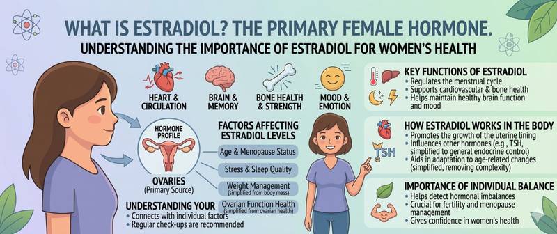
Estradiol is called the primary female hormone — and for good reason. But behind this familiar label lies a substance with a far broader role: it governs not only the menstrual cycle and fertility, but also bone density, vascular health, mood, and even the lipid profile of the blood. Let's look at what estradiol actually is, why its normal values change every week of the menstrual cycle, and what elevated or low levels mean.
What Is Estradiol and How It Differs From Estrogen
"Estrogen" is a collective term for a group of hormones. The human body produces three: estrone (E1), estradiol (E2), and estriol (E3). Estradiol is the most potent of the three — 10 to 80 times more active than estrone — and it is the dominant form during reproductive years.
In women, estradiol is produced primarily in the ovaries (granulosa cells of follicles); in men, in the testes and through peripheral conversion of testosterone in adipose tissue. Small amounts are produced by the adrenal glands in both sexes.
Estradiol receptors are present in virtually every tissue: uterus, breasts, bones, blood vessels, brain, skin, intestines. This is why changes in estradiol levels are felt across many systems simultaneously — from mood to joint health.
The Role of Estradiol in the Body
Reproductive function. Estradiol drives follicle growth, prepares the endometrium for implantation, and regulates ovulation through feedback with the pituitary gland (an estradiol surge triggers the LH peak). Without adequate estradiol, a normal cycle is not possible.
Bone tissue. Estradiol inhibits bone resorption by osteoclasts. After menopause, when its level falls, bone breakdown accelerates faster than it can be rebuilt — this is the foundation of postmenopausal osteoporosis. Adequate calcium, vitamin D, and estradiol are all required for normal bone mineralisation — all three are interconnected.
Cardiovascular system. Estradiol protects the vascular endothelium, raises HDL, and lowers LDL. This explains why women before menopause have significantly lower coronary artery disease risk than men of the same age — and why that risk rises sharply after menopause.
Central nervous system. Estradiol influences serotonin and dopamine synthesis. Premenstrual syndrome, postpartum depression, and mood instability in perimenopause are largely consequences of estradiol fluctuations.
In men. Normal estradiol is necessary for libido, erectile function, bone density, and normal spermatogenesis. Too low estradiol in men is as much a problem as too high.
Normal Estradiol Values in Women by Cycle Phase
The key feature of estradiol interpretation in women of reproductive age: normal values differ fundamentally by cycle phase. Evaluating the result without specifying the cycle day is meaningless.
| Cycle phase | Normal estradiol (pmol/L) | Normal (pg/mL) |
|---|---|---|
| Follicular (days 1–13) | 68–1270 | 18–147 |
| Ovulatory peak (day 14) | 131–1655 | 36–450 |
| Luteal (days 15–28) | 91–861 | 25–235 |
| Postmenopause | < 73 | < 20 |
| Pregnancy 1st trimester | 215–4300 | 58–1170 |
During pregnancy, estradiol is produced by the placenta — levels rise hundreds of times above non-pregnant norms.
Normal Estradiol Values in Men
| Group | Normal (pmol/L) | Normal (pg/mL) |
|---|---|---|
| Adult men | 40–161 | 11–44 |
In men, elevated estradiol is most commonly linked to obesity (adipose tissue converts testosterone to estradiol) or anabolic steroid use.
When to Test Estradiol
For women of reproductive age: on days 2–5 of the cycle (early follicular phase), when the goal is to assess basal levels and ovarian function. To assess ovulation — around the middle of the cycle (days 12–14). Always fast before the draw.
For postmenopausal women and men — any day, morning, fasting.
Always note the cycle day on the test request — without it, the laboratory cannot apply the correct reference range.
Causes of Elevated Estradiol
In women: estrogen-producing ovarian tumours; endometriosis — ectopic endometrial tissue produces estradiol locally; obesity — adipose tissue produces estrone, which partially converts to estradiol; liver cirrhosis — impaired estrogen metabolism; use of estrogen-containing medications (combined oral contraceptives, HRT); physiological pre-ovulatory surge — not pathological.
In men: obesity (the main cause); testicular tumours; liver cirrhosis; primary hypogonadism with elevated FSH and LH.
Causes of Low Estradiol
In women: primary ovarian insufficiency (premature menopause); hypogonadotropic hypogonadism — impaired pituitary signalling; anorexia nervosa or rapid weight loss — adipose tissue is needed for estrogen synthesis; intense athletic training (athletic amenorrhoea); hyperprolactinaemia — excess prolactin suppresses estradiol production; hypothyroidism — thyroid dysfunction disrupts hormonal balance; perimenopause and menopause — physiological decline of ovarian function.
In men: primary hypogonadism; use of aromatase inhibitors.
Estradiol and PCOS
In polycystic ovary syndrome, estradiol is often normal or mildly elevated — but the pattern is disrupted: many small follicles, none dominant, ovulation does not occur. Testosterone and LH are elevated, and the LH/FSH ratio exceeds 2. Estradiol in this context is just one of several markers; PCOS diagnosis requires a combination of criteria.
Estradiol and Menopause
The decline of estradiol in perimenopause is gradual but systemic. Hot flushes, sleep disruption, mucosal dryness, accelerated bone loss, and a shift in the lipid profile toward atherogenicity — all are direct consequences of estradiol deficiency. Menopausal hormone therapy (MHT) is directed specifically at replenishing estradiol.
When to See a Doctor
Routine referral to a gynaecological endocrinologist for: menstrual irregularities (amenorrhoea, oligomenorrhoea) lasting more than 3 months; infertility; symptoms of early menopause before age 40; gynaecomastia in men; symptoms of estrogen deficiency (hot flushes, vaginal dryness, osteoporosis at a young age).
Summary
Estradiol has a remarkably wide spectrum of action that extends far beyond the reproductive system. Its level must always be assessed in the context of the cycle phase in women, and alongside other hormones — FSH, LH, prolactin, testosterone. An isolated number without clinical context tells almost nothing. Long-term estradiol deficiency affects bones, blood vessels, and the brain — it is not "just a hormonal imbalance."
This article is for informational purposes only. Interpretation of test results and treatment decisions are the responsibility of a physician.
For informational purposes only
This article is for informational purposes only and does not constitute medical advice, diagnosis, or treatment. Please consult a healthcare professional for medical guidance.