Pancreatic Cancer: Symptoms, Diagnosis and Treatment Guide
Reviewed by the LabReadAI medical team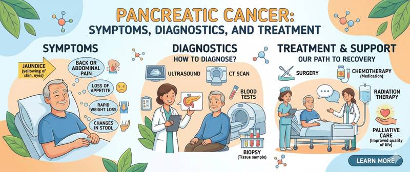
The skin turns yellow. A dull ache in the upper abdomen begins radiating into the back. Weight is dropping for no apparent reason and appetite has been absent for weeks. This is how it typically begins — not dramatically, but gradually and without obvious cause. Pancreatic cancer is found at a surgically resectable stage in only 15–20% of patients: the organ sits deep in the retroperitoneum, and by the time symptoms emerge the tumour has usually already spread beyond it.
What Pancreatic Cancer Is and Its Types
Pancreatic cancer is a malignant tumour arising from cells of the pancreas. In 90–95% of cases it is pancreatic ductal adenocarcinoma (PDAC), a tumour of the duct-lining epithelium. It is one of the most aggressive malignancies known: five-year overall survival across all stages is approximately 11–12%.
The anatomical location of the tumour determines the clinical picture. Cancer of the pancreatic head (60–70% of cases) compresses the common bile duct early, producing the characteristic painless obstructive jaundice. Cancer of the body and tail (30–40%) grows silently for much longer: the bile duct is not involved, there is no jaundice, and symptoms appear only when the tumour invades adjacent organs or encases major vessels.
Rare forms include pancreatic neuroendocrine tumours (pNETs, ~5%) — significantly less aggressive, often hormonally active (insulinoma, gastrinoma, glucagonoma). Their markers and treatment are fundamentally different from PDAC.
TNM/AJCC staging:
| Stage | Description | 5-year survival |
|---|---|---|
| I | Tumour confined to the pancreas | 20–25% |
| II | Local spread without major vessel involvement | 8–12% |
| III | Involvement of the coeliac axis or superior mesenteric artery | 3–5% |
| IV | Distant metastases (liver, peritoneum, lungs) | 1–3% |
Approximately 80–85% of patients present at stage III or IV. This is the principal cause of the dismal prognosis: not the biology of the tumour alone, but chronically lost time.
Causes and Risk Factors
Smoking — the most significant modifiable factor, accounting for 20–30% of all cases. Risk in smokers is approximately double that of non-smokers; it approaches population risk within 10 years of cessation.
Chronic pancreatitis — one of the strongest risk factors: risk is 10–20 times higher than in the general population. The mechanism is chronic inflammation with cellular damage and accumulation of oncogenic mutations. Risk is particularly high in hereditary pancreatitis (PRSS1 gene mutations).
Type 2 diabetes approximately doubles the risk. There is an important bidirectional signal: newly diagnosed diabetes in a person over 50 who is not overweight and has no family history of diabetes may be paraneoplastic — the tumour destroys the insulin-producing islet cells. New-onset diabetes in an "atypical" patient is a reason to exclude pancreatic cancer.
Obesity raises risk by 20–40%, particularly in combination with a sedentary lifestyle. Visceral fat drives chronic inflammation and hyperinsulinaemia.
Age: 90% of cases are diagnosed after age 55; peak incidence is at 65–75 years.
Alcohol: heavy use increases risk through acute pancreatitis evolving into chronic disease.
Hereditary factors account for 5–10% of cases:
- BRCA2 and PALB2 mutations — 3–6 times higher risk; especially important when family history includes breast and ovarian cancer
- ATM mutations — 4–5 times higher risk
- Lynch syndrome (MMR genes) — 8–9 times higher risk
- Peutz–Jeghers syndrome (STK11) — risk approximately 130 times higher
- FAMMM syndrome (CDKN2A) — principally associated with melanoma but carries 10–20 times elevated pancreatic cancer risk
Symptoms: Why Pancreatic Cancer Is Diagnosed So Late
The pancreas is a "silent" organ: it has no pain receptors on its surface, lies behind the stomach and intestinal loops, and is separated from the body surface by several centimetres of tissue. A small tumour there can grow for years without producing any symptoms.
Symptoms of head of pancreas cancer:
- Jaundice — yellowing of the skin and sclerae, dark urine, pale stools. This is not inflammatory jaundice but painless mechanical obstruction: bile cannot drain into the intestine because the bile duct is compressed by the tumour. The painless character of this jaundice distinguishes it from gallstone-related obstruction, where pain almost invariably accompanies or precedes the yellowing
- Pruritus — caused by accumulation of bile salts in the skin during cholestasis
- Pain in the right upper quadrant or epigastrium
Symptoms of body and tail cancer:
- Dull aching pain in the upper abdomen radiating to the back and lower spine. A characteristic feature: pain worsens when lying supine and eases when leaning forward — the tumour is pressing on the coeliac plexus
- New or dramatically worsened diabetes without an obvious cause
General symptoms (any location):
- Unexplained weight loss — often the first and most noticeable symptom: 5–10% of body weight over 3–6 months with no clear explanation
- Marked loss of appetite, aversion to fatty food
- Steatorrhoea — greasy, floating, foul-smelling stools from exocrine pancreatic insufficiency
- Migratory superficial thrombophlebitis (Trousseau's syndrome) — recurrent painful vein inflammation at different sites without an obvious cause; a paraneoplastic syndrome particularly characteristic of pancreatic cancer
- Depression and anxiety — documented to precede the diagnosis by several months through a mechanism that remains incompletely understood
Incidental detection: in approximately 10–15% of patients the tumour is found incidentally during CT or ultrasound performed for another reason — the only realistic route to early-stage detection in the absence of a general screening programme.
Diagnosis: CT, Endosonography and Laboratory Tests
Diagnosis rests on the combination of imaging and laboratory data. Morphological confirmation is mandatory before any treatment is started.
Multiphase pancreatic-protocol CT — the investigation of choice for initial staging. Triphasic contrast enhancement visualises the tumour, assesses its relationship to the superior mesenteric artery, coeliac axis and portal vein (the key determinant of resectability), and detects hepatic metastases and enlarged lymph nodes.
Endoscopic ultrasound (EUS) — the most sensitive method for small tumours (< 2 cm). The probe is positioned immediately adjacent to the pancreas in the stomach or duodenum, eliminating the distance problem. EUS also permits simultaneous fine-needle aspiration biopsy for histological confirmation.
ERCP (endoscopic retrograde cholangiopancreatography) — for obstructive jaundice: permits biliary stenting and cytological sampling.
MRI with MRCP — for detailed assessment of biliary and pancreatic duct anatomy and characterisation of cystic lesions.
Laboratory tests:
Complete blood count — normochromic normocytic anaemia in chronic disease; thrombocytosis as a paraneoplastic phenomenon driven by tumour-derived thrombopoietic cytokines.
Tumour marker panel — CA 19-9 and CEA; interpreted only after imaging has established the diagnosis.
Liver biochemistry — bilirubin (total and direct), alkaline phosphatase, GGT, ALT, AST: a cholestatic pattern confirms bile duct obstruction. LDH rises with extensive hepatic metastatic involvement.
Pancreatic panel — amylase and lipase: in pancreatic cancer these are often normal or only mildly elevated; significant elevation points more to obstructive pancreatitis than to the tumour itself.
Glucose and HbA1c — when new-onset diabetes may be paraneoplastic.
Tumour Markers: CA 19-9 and Its Role
CA 19-9 (carbohydrate antigen 19-9) — the primary tumour marker for pancreatic cancer. Normal range < 37 U/mL. Sensitivity for PDAC: 70–80%; specificity: 80–90%. It is the best available marker, but its limitations are clinically important.
A biological limitation: 5–10% of people are genetically unable to produce CA 19-9 — they are Lewis-antigen-negative. In these individuals the marker remains at or near zero even in advanced disease. This is not reassuring — it simply reflects a biological characteristic that must be accounted for in interpretation.
False-positive elevation of CA 19-9 occurs with any cause of obstructive jaundice (bile acids stimulate its synthesis), chronic pancreatitis, liver cirrhosis, gastric cancer and colorectal cancer. A high CA 19-9 in a jaundiced patient does not confirm pancreatic cancer — jaundice itself raises it. Reassessing after biliary drainage and bilirubin normalisation is a mandatory step before clinical conclusions are drawn.
Clinical applications of CA 19-9:
- Post-operative monitoring: normalisation within 3–4 weeks is a favourable sign; persistent elevation suggests incomplete resection or occult metastases
- Chemotherapy response: a fall of 50–75% after 2–3 cycles indicates response; a rise during treatment signals progression
- Early relapse detection: rising CA 19-9 after remission often precedes CT evidence of recurrence by 4–6 weeks
- CA 19-9 is not used for primary diagnosis: no tumour marker replaces biopsy
Treatment of Pancreatic Cancer
Treatment strategy is determined by resectability — the central concept in pancreatic surgery, assessed by the tumour's relationship to the superior mesenteric artery, coeliac axis and portal vein.
Surgery — the only potentially curative option; applicable to 15–20% of patients at presentation.
The Whipple procedure (pancreaticoduodenectomy) is performed for cancer of the head: it removes the pancreatic head, duodenum, gallbladder, common bile duct, part of the stomach and regional lymph nodes. This is one of the most technically demanding operations in abdominal surgery; in specialist centres, operative mortality is below 3%.
Distal pancreatectomy — for body and tail cancers: the left half of the pancreas is removed together with the spleen.
After surgery — adjuvant chemotherapy for 6 months: the mFOLFIRINOX regimen (oxaliplatin + irinotecan + 5-FU) reduces recurrence risk by 35–40% compared with gemcitabine monotherapy.
Borderline resectable disease: the tumour abuts but does not circumferentially encase major vessels. Strategy: upfront chemotherapy, then CT reassessment. Approximately 30–40% of such patients achieve resectability.
Locally advanced (unresectable) cancer (stage III): FOLFIRINOX ± radiotherapy. Median survival 12–15 months; secondary resection is achievable in a subset of responders.
Metastatic cancer (stage IV):
- Good performance status (ECOG 0–1): mFOLFIRINOX — median overall survival 11–12 months
- Frail patients: gemcitabine + nab-paclitaxel — median 8–9 months
- BRCA1/2 or PALB2 mutations: platinum-based regimens followed by maintenance olaparib; key trials showed this approximately doubles progression-free survival
- MSI-H tumours (~1% of PDAC): pembrolizumab
Palliative interventions:
- Biliary stenting — relieves obstructive jaundice in inoperable head cancers
- Coeliac plexus block — highly effective pain management when the tumour invades the retroperitoneal space
- Pancreatic enzyme replacement therapy (PERT) — corrects exocrine insufficiency, improves nutritional status and quality of life
When to See a Doctor and Prevention
Consult a doctor (gastroenterologist or surgeon) promptly when one or more of the following apply:
- painless yellowing of the skin or whites of the eyes;
- dull back pain in the upper spine not related to movement or posture;
- unexplained weight loss of 5% or more over 2–3 months;
- newly diagnosed or dramatically worsening diabetes in someone over 50 without obesity or family history;
- recurrent episodes of superficial vein inflammation at different body sites;
- persistent loss of appetite lasting more than 3 weeks.
Screening in the general population is not currently recommended. For high-risk groups (Lynch syndrome, BRCA2 mutation, FAMMM, Peutz–Jeghers syndrome, hereditary chronic pancreatitis), surveillance protocols exist: annual EUS plus MRI/MRCP beginning at age 40 or 10 years before the youngest affected family member's diagnosis.
Prevention: smoking cessation is the single evidence-based strategy that measurably reduces population risk. Weight management, alcohol limitation and timely treatment of chronic pancreatitis offer additional risk reduction.
Pancreatic cancer remains one of the most challenging diagnoses in oncology. But when identified at a resectable stage, long-term survival is genuinely achievable. The only path to that outcome is not dismissing symptoms and seeking evaluation without delay.
This content is for informational purposes only and does not replace professional medical advice.
For informational purposes only
This article is for informational purposes only and does not constitute medical advice, diagnosis, or treatment. Please consult a healthcare professional for medical guidance.