Nephrotic Syndrome: Symptoms, Causes and Treatment
Reviewed by the LabReadAI medical team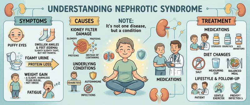
Oedema that starts with puffy eyes in the morning and gradually spreads to the whole body, together with frothy urine and profound fatigue — this is the classic presentation of nephrotic syndrome. Behind this syndrome lies one fundamental disruption: the kidney filter has become permeable to protein. Everything else follows from that.
What Nephrotic Syndrome Is
Nephrotic syndrome is a clinico-laboratory complex defined by four classic features:
- Massive proteinuria — urinary protein loss > 3.5 g/day in adults (> 40 mg/m²/hour in children). This is the defining, primary feature — all others derive from it.
- Hypoalbuminaemia — blood albumin < 35 g/L (often < 25 g/L in fully developed syndrome).
- Generalised oedema — soft, symmetrical, beginning periorbital, then spreading to ankles, legs, scrotum, ascites, pleural effusion.
- Hyperlipidaemia — elevated total cholesterol, LDL, and triglycerides. The liver's compensatory response to falling oncotic pressure.
A fifth feature often included: lipiduria — fatty casts and "Maltese crosses" in urine sediment under polarised light.
Nephrotic syndrome is not a standalone diagnosis — it is always the expression of an underlying disease of the glomerular apparatus of the kidneys.
Mechanism: Why Protein Is Lost
The glomerular filter normally bars large molecules — albumin (69 kDa) is retained at three levels: the endothelium, the basement membrane, and podocytes (cells with "foot processes" covering the outer surface of the basement membrane). In nephrotic syndrome, podocytes are damaged — their foot processes flatten or fuse. The filter becomes permeable to albumin and other proteins.
Consequences of massive proteinuria:
- Fall in plasma oncotic pressure → fluid shifts from vessels into tissues → oedema
- Hypovolaemia → activation of the renin-angiotensin-aldosterone system → sodium and water retention → worsening oedema
- Protein deficit signal → liver compensatorily increases synthesis of all proteins including lipoproteins → hyperlipidaemia
- Loss of anticoagulant proteins (antithrombin III, protein C and S) → hypercoagulability — thrombosis risk
Causes of Nephrotic Syndrome
In children
Minimal change disease (MCD) — the cause of 90% of nephrotic syndrome in children under 8. Light microscopy shows no changes — only electron microscopy reveals podocyte foot process fusion. Responds excellently to glucocorticoids: remission in 90% of children. Prognosis is generally very good.
In adults
Primary (idiopathic) glomerulopathies:
- Minimal change disease (10–15%) — in adults often associated with Hodgkin's lymphoma and NSAIDs
- Focal segmental glomerulosclerosis (FSGS) — the most common primary cause in US adults; frequently treatment-resistant
- Membranous nephropathy — the leading primary cause in White adults; idiopathic (anti-PLA2R antibodies) or secondary
- Membranoproliferative glomerulonephritis
Secondary (systemic disease):
- Diabetes mellitus — diabetic nephropathy: the most common cause of nephrotic syndrome in adults globally
- Amyloidosis — amyloid fibril deposition in glomeruli; systemic AL-amyloidosis or reactive AA-amyloidosis
- Systemic lupus erythematosus (SLE) — lupus nephritis class V
- Infections — hepatitis B (membranous nephropathy), hepatitis C (MPGN), HIV (FSGS), syphilis, malaria
- Drugs — NSAIDs, gold salts, penicillamine, captopril, heroin
Nephrotic Syndrome Symptoms: Oedema, Proteinuria and Complications
Oedema — the hallmark of nephrotic syndrome. Unlike cardiac oedema (starting inferiorly — at the ankles), nephrotic oedema characteristically begins with the face: periorbital puffiness on waking is one of the earliest signs. Over time, oedema generalises: legs, scrotum, ascites, hydrothorax. The overlying skin is pale, soft, and pitting.
Frothy urine — the excess protein creates foam that persists long after the stream ends. One of the first symptoms patients notice.
General weakness and fatigue — consequence of hypoalbuminaemia and anaemia (urinary loss of transferrin and erythropoietin).
Hyperlipidaemia and xanthomas — with prolonged nephrotic syndrome, xanthoma deposits may appear on the skin.
Complications:
- Thrombosis and thromboembolism — renal vein thrombosis (classic complication of membranous nephropathy), deep vein thrombosis, pulmonary embolism. Driven by urinary loss of anticoagulant proteins and hyperfibrinogenaemia.
- Infections — loss of immunoglobulins and complement components → high susceptibility to encapsulated bacteria (pneumococcus, Haemophilus). Pneumococcal peritonitis is characteristic in children with MCD.
- Acute kidney injury — in 25–30% of patients; mechanisms: hypovolaemia, renal vein thrombosis, nephrotoxic drugs.
- Atherosclerosis — chronic hyperlipidaemia with prolonged nephrotic syndrome accelerates arterial disease.
Diagnosis: Key Lab Tests
Urine:
- 24-hour proteinuria > 3.5 g, or urine protein-to-creatinine ratio > 3.5 g/g — the diagnostic criterion
- Urinalysis: protein +++, fatty casts, "Maltese crosses" under polarised light
- Complete blood count with differential
Blood:
- Albumin — reduced (< 35 g/L); severity of hypoalbuminaemia correlates with syndrome severity
- Total protein — reduced
- Total cholesterol, LDL, triglycerides — elevated
- Creatinine, urea — kidney function assessment
- Coagulation panel — assess hypercoagulability
- C-reactive protein, ESR — inflammatory markers
Aetiological workup:
- Anti-dsDNA, ANA — SLE
- HBsAg, HCV, HIV
- Anti-PLA2R antibodies — membranous nephropathy
- Bence-Jones protein in urine, immunofixation — amyloidosis, multiple myeloma
- Kidney biopsy — the primary method of morphological diagnosis in adults
Treatment of Nephrotic Syndrome
Treatment is two-pronged: specific therapy for the underlying disease + symptomatic management.
Specific therapy:
- Minimal change disease: glucocorticoids (prednisolone 1 mg/kg/day) — remission in 80–90% of children and ~75% of adults. For relapses — tacrolimus, cyclophosphamide, rituximab.
- Membranous nephropathy: 30% undergo spontaneous remission. For progression — Ponticelli regimen (corticosteroids + chlorambucil), tacrolimus, rituximab.
- FSGS: glucocorticoids — first line; for resistance — calcineurin inhibitors.
- Diabetic nephropathy: RAAS inhibitors (ACEi/ARB) — reduce proteinuria; SGLT2 inhibitors — nephroprotective effect; glycaemic control (target HbA1c).
Symptomatic management:
- Oedema: sodium restriction (< 2 g/day), loop diuretics (furosemide). With hypovolaemia — use cautiously; risk of AKI.
- Hyperlipidaemia: statins — reduce cardiovascular risk in prolonged syndrome.
- Thrombosis prevention: at albumin < 20–25 g/L in membranous nephropathy — anticoagulant prophylaxis is considered.
- Dietary protein: moderate intake (0.8–1.0 g/kg/day) — high protein intake worsens proteinuria.
- Vaccination: pneumococcal and influenza vaccines — with prolonged nephrotic syndrome.
When to Seek Urgent Medical Attention
- Newly appeared generalised oedema + frothy urine — nephrology referral within days
- Dyspnoea with oedema — possible hydrothorax or pulmonary embolism
- Leg pain + swelling + redness — deep vein thrombosis; call emergency services
- Flank pain + blood in urine + worsening oedema — possible renal vein thrombosis
- Fever + abdominal pain in a child with nephrotic syndrome — rule out spontaneous peritonitis
This article is for informational purposes only and does not replace consultation with a qualified nephrologist.
For informational purposes only
This article is for informational purposes only and does not constitute medical advice, diagnosis, or treatment. Please consult a healthcare professional for medical guidance.