Sleep Apnoea: Causes, Symptoms, Diagnosis and Treatment
Reviewed by the LabReadAI medical team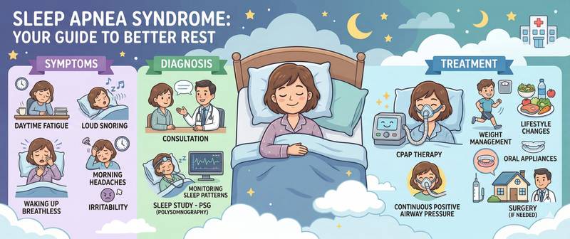
Your partner says you snore loudly and sometimes stop breathing altogether. You wake up feeling as though you haven't slept at all. You nod off during the day despite spending eight hours in bed. These are the classic symptoms of sleep apnoea — one of the most common and most underdiagnosed conditions in medicine, affecting 15–30% of men and 10–15% of women, while the vast majority remain unaware of their diagnosis.
What Is Sleep Apnoea and Its Types
Obstructive sleep apnoea (OSA) is a sleep-disordered breathing condition characterised by repeated episodes of partial or complete upper airway obstruction lasting at least 10 seconds, accompanied by falls in blood oxygen saturation and sleep fragmentation.
The mechanism of obstructive apnoea: during sleep, the pharyngeal muscles relax, the soft palate and tongue base drop and occlude the airway. Blood oxygen falls, the brain registers the threat and "wakes" the person for a fraction of a second — enough for the muscles to re-tense and breathing to resume. The person rarely remembers these arousals, yet there may be dozens or hundreds per night. This is why sufferers wake feeling unrefreshed regardless of how long they spent in bed.
Severity is graded by the apnoea-hypopnoea index (AHI) — the number of breathing cessations and reductions per hour of sleep:
| Severity | AHI (events/hour) |
|---|---|
| Mild | 5–14 |
| Moderate | 15–29 |
| Severe | ≥ 30 |
Beyond obstructive apnoea, a central form (CSA) exists — respiratory efforts cease completely due to disrupted central respiratory drive. CSA is less common and typically associated with heart failure or post-stroke status. A mixed form combines both mechanisms.
Causes and Risk Factors
Obesity — the leading and most prevalent risk factor. Obesity is present in 60–70% of OSA patients: excess adipose tissue in the neck (circumference > 43 cm in men, > 38 cm in women) directly narrows the pharyngeal lumen, while chest and abdominal fat further restricts diaphragmatic excursion.
Anatomical features: retrognathia (recessed jaw), tonsillar and adenoid hypertrophy, elongated uvula, and deviated nasal septum. This is why OSA frequently occurs in lean individuals with specific craniofacial anatomy.
Sex and age: men are two to three times more commonly affected than women of reproductive age. After menopause, the gap narrows — progesterone, which maintains pharyngeal muscle tone, is no longer produced. After age 50, OSA prevalence increases sharply in both sexes.
Alcohol and sedatives — worsen apnoea: they relax pharyngeal muscles and suppress the arousal response.
Smoking — chronic upper airway mucosal inflammation increases pharyngeal collapsibility.
Body position: most patients have worse apnoea when sleeping supine — gravity displaces the tongue and soft tissue posteriorly.
Hypothyroidism — one of the most common endocrine contributors; it reduces pharyngeal muscle tone and causes soft tissue oedema. This is why TSH is checked in all patients with newly diagnosed OSA.
Symptoms of Sleep Apnoea: Night and Day
OSA symptoms divide into those occurring at night (witnessed by a partner or recorded by devices) and those experienced during the day.
Night-time symptoms:
- Loud, intermittent snoring with silent pauses (breathing cessations) — the most characteristic sign
- Witnessed apnoeas followed by a loud gasp or snort as breathing resumes
- Restless sleep with frequent position changes
- Night sweats
- Nocturia — frequent night-time urination (caused by increased intrathoracic pressure and atrial natriuretic peptide release)
- Night-time headaches, choking sensations, awakenings
Daytime symptoms:
- Excessive daytime sleepiness — the central symptom. Patients fall asleep while driving, in meetings, watching television. Epworth Sleepiness Scale score > 10 is clinically significant
- Morning headache — from hypercapnia (CO₂ accumulation) during nocturnal apnoeas
- Impaired concentration and memory
- Irritability, depression, emotional lability
- Reduced libido and erectile dysfunction in men
Important: many patients adapt to chronic sleepiness and stop perceiving it as abnormal, considering their state "normal". Systematic questioning about apnoea symptoms is therefore more productive than waiting for spontaneous complaints.
Diagnosing Sleep Apnoea
OSA diagnosis is based on objective sleep monitoring — subjective symptoms and sleepiness scales merely justify referral but do not confirm the diagnosis.
Polysomnography (PSG) — the gold standard. Overnight monitoring in a sleep laboratory records simultaneously: EEG (sleep stages), EOG (eye movements), chin EMG (muscle tone), ECG, respiratory effort (thoracic and abdominal bands), oronasal airflow, pulse oximetry and body position. It provides a complete picture of all breathing disorders and sleep architecture.
Ambulatory polygraphy (home sleep testing) — a simplified PSG without EEG. The patient wears a portable device at home. Adequate for confirming moderate and severe OSA but may underestimate severity in mild disease. More accessible and convenient — used as the initial test when pre-test probability is high.
Epworth Sleepiness Scale (ESS) — an eight-item self-questionnaire rating the likelihood of dozing in different situations. ESS > 10 is an indication for diagnostic evaluation.
ENT examination — mandatory to identify anatomical factors (tonsillar hypertrophy, septal deviation) that influence treatment selection.
What Blood Tests Are Ordered in Sleep Apnoea
No laboratory tests are specific to OSA. However, several tests are mandatory in initial evaluation — they identify causes and complications.
- TSH — exclude hypothyroidism as a cause of apnoea and daytime sleepiness
- Haemoglobin and haematocrit — secondary polycythaemia (elevated haemoglobin as a response to chronic hypoxia) occurs in severe untreated OSA; an unexpectedly high haemoglobin is itself a reason to screen for OSA
- Metabolic syndrome assessment — glucose, lipid profile, blood pressure: OSA and metabolic syndrome are tightly clustered, each worsening the other
- Insulin resistance markers — chronic nocturnal hypoxia in OSA reduces insulin sensitivity independently of obesity
Overnight pulse oximetry — a simple home monitoring of blood oxygen saturation — does not replace PSG but provides supporting evidence when OSA is suspected: desaturation below 90% during sleep is a characteristic pattern.
Sleep Apnoea and Cardiovascular Disease
OSA is not merely a "sleep problem" — it is a systemic condition with far-reaching cardiovascular consequences.
Arterial hypertension is present in 50% of OSA patients. Mechanism: repetitive apnoeas activate the sympathetic nervous system and RAAS, sustaining elevated blood pressure. A characteristic pattern in OSA-related hypertension is nocturnal and morning elevation with absent physiological overnight dipping ("non-dipper" pattern). OSA is the most common cause of resistant hypertension.
Heart failure — OSA is present in 30–40% of patients with chronic HF. Apnoea worsens heart failure through nocturnal hypoxia, sympathetic activation and increased left ventricular afterload. Treating OSA in HF patients improves ejection fraction and reduces hospitalisations.
Atherosclerosis — chronic nocturnal hypoxia and oxidative stress accelerate coronary and carotid atherosclerotic plaque development. The risk of myocardial infarction and stroke with severe untreated OSA is two to three times higher than background risk.
Type 2 diabetes — OSA is an independent risk factor. Nocturnal hypoxia impairs insulin secretion and elevates cortisol, independently worsening glycaemic control.
Atrial fibrillation — risk is two to four times higher in OSA. Apnoea causes mechanical atrial stretch, sympathetic activation and conditions conducive to arrhythmia development.
Treatment: CPAP Therapy and Other Approaches
Treatment of OSA depends on severity, dominant contributing factors and co-existing conditions.
Continuous positive airway pressure (CPAP) therapy — the gold standard for moderate and severe OSA. The device delivers air at constant pressure through a mask, preventing pharyngeal collapse. With regular use (≥ 4 hours per night):
- elimination of snoring and apnoeas in 90–95% of patients;
- reduction in daytime sleepiness, often from the very first night;
- systolic blood pressure reduction of 2–7 mmHg in resistant hypertension;
- reduced cardiovascular event risk with long-term use.
The main challenge with CPAP is adherence: approximately 30–50% of patients discontinue treatment in the first months due to mask discomfort or device noise. Mask fitting, regular follow-up and specialist support are critical for success.
Weight loss — a 10% weight reduction decreases AHI by approximately 26% on average. In patients who achieve significant weight loss through bariatric surgery, OSA resolves entirely in 40–80%.
Positional therapy — for position-dependent OSA (worse supine): devices or positional pillows preventing supine sleep. Effective in mild to moderate positional OSA.
Mandibular advancement devices (MAD) — oral appliances that advance the lower jaw forward. An alternative to CPAP in mild to moderate OSA; less effective on average but better tolerated by some patients.
Surgical treatment — tonsillectomy for tonsillar hypertrophy (particularly in children), uvulopalatopharyngoplasty, nasal septal surgery. Surgery is less predictable than CPAP and applied for specific anatomical indications.
Hypoglossal nerve stimulation — a modern option for CPAP-intolerant patients: an implanted neurostimulator synchronised with breathing activates the tongue muscles at each inhale.
When to See a Doctor
See a sleep medicine specialist, pulmonologist or ENT physician when two or more of the following apply:
- loud snoring witnessed by a bed partner;
- observed breathing pauses during sleep;
- excessive daytime sleepiness despite adequate time in bed;
- recurring morning headaches;
- hypertension not responding to standard medication;
- obesity with neck circumference > 43 cm in men or > 38 cm in women.
Untreated apnoea gradually and silently damages health: it raises blood pressure, overloads the heart and disrupts metabolic balance. At the same time, CPAP therapy often transforms a patient's life after a single night of use. Do not postpone seeing a doctor.
This content is for informational purposes only and does not replace professional medical advice.
For informational purposes only
This article is for informational purposes only and does not constitute medical advice, diagnosis, or treatment. Please consult a healthcare professional for medical guidance.