High and Low Hematocrit and Red Blood Cell Count: Causes
Reviewed by the LabReadAI medical team
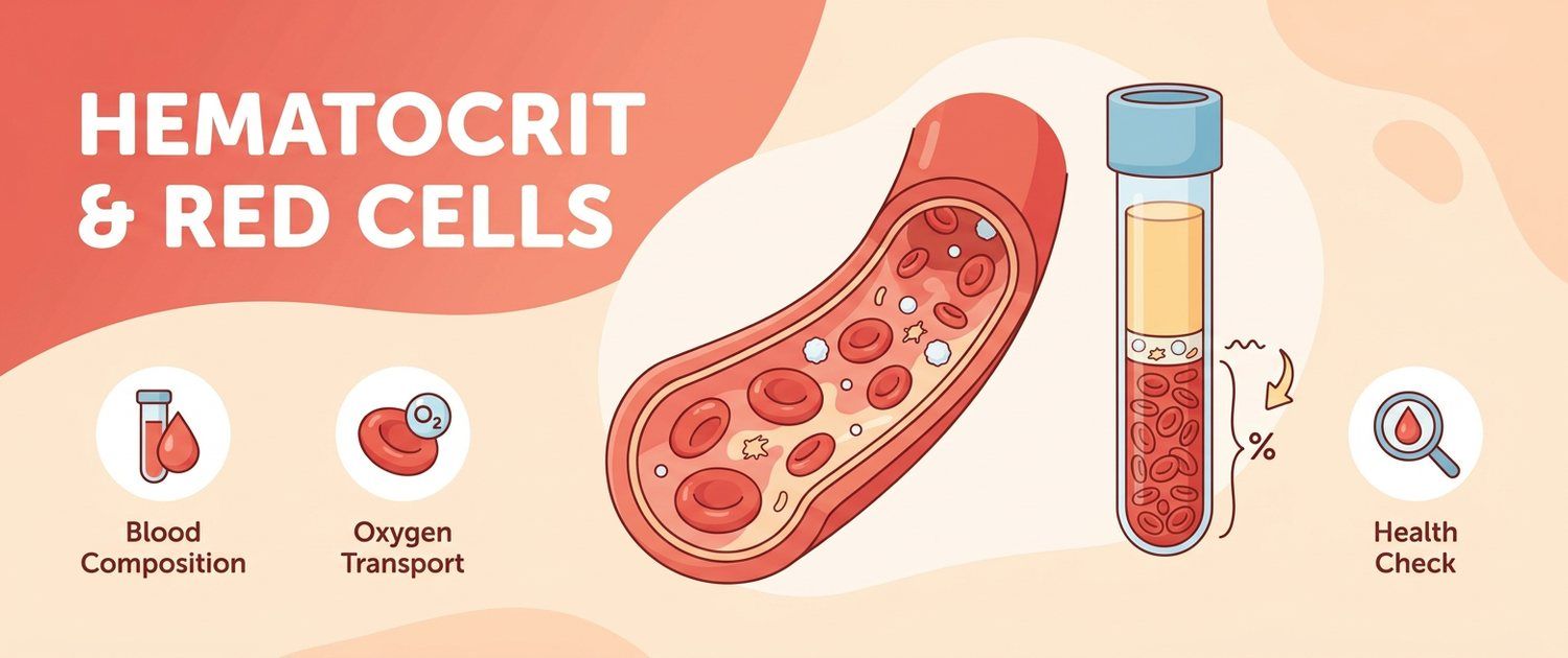
Red cells and hematocrit sit next to each other on a report and almost always move together — they are two records of the same thing. Red cells are counted in units (× 10¹²/L); hematocrit expresses the same mass as a share of volume (%). Reading a full report is easier with the general guide: Blood Test Guide.
The key to the whole subject is the denominator. Hematocrit is the volume of red cells divided by the volume of whole blood. The numerator changes slowly: a red cell lives about 120 days and the marrow needs weeks to alter their number appreciably. The denominator — plasma volume — changes within hours. Hence the practical rule: a fast jump in hematocrit is almost always about water, not about cells.
Below: why a high hematocrit is most often dehydration, when a red blood cell count truly rises because of disease, what the normal range is in men, women and children, and what low values mean.
Normal red blood cell count and hematocrit in men, women and children
Reference intervals differ by sex: testosterone stimulates red cell production, so the male range sits higher.
| Group | Red cells, × 10¹²/L | Hematocrit, % |
|---|---|---|
| Men | 4.3–5.7 | 39–49 |
| Women | 3.8–5.1 | 35–45 |
| Pregnancy, third trimester | 3.5–4.8 | 31–41 |
| Newborn | 4.0–6.6 | 45–65 |
| 1–6 months | 3.1–4.5 | 29–41 |
| 1–6 years | 3.7–4.9 | 32–42 |
| 6–12 years | 4.0–5.2 | 34–43 |
| 12–18 years | 4.1–5.5 | 35–47 |
Two things deserve note. First, in newborns the hematocrit is physiologically very high and halves over the first months — that is normal, not developing anaemia. Second, in pregnancy the lower limit is honestly below the female range, because plasma volume grows by 40–50% by the third trimester while red cell mass grows only 20–30%. These age and physiological shifts are covered in CBC with Differential and CBC in Pregnancy.
High hematocrit: dehydration and relative erythrocytosis
Before disease is considered, the commonest cause is ruled out. Relative erythrocytosis means exactly as many cells as before, in less plasma. Hematocrit rises, the red blood cell count "rises", haemoglobin rises too — and none of it reflects the marrow.
What shrinks plasma volume:
- Not drinking enough, especially in heat and during exertion.
- Vomiting, diarrhoea, fever, heavy sweating.
- Diuretic drugs.
- Burns and other large fluid losses.
- Giving blood in the morning after a long night without water — the most ordinary version of all.
How relative erythrocytosis is recognised: the number is high, but all three red-cell values rose proportionally, the indices are unchanged, the person feels normal, and a repeat after normal fluid intake returns to baseline. Haemoconcentration nominally lifts other lines too — the same effect is discussed in High and Low Platelets and High and Low Lymphocytes.
A separate version is the steady mild elevation seen with excess weight, high blood pressure and regular alcohol, where plasma volume is chronically reduced. Hematocrit sits near 50–52%, does not climb over years and comes with no change in other lineages.
High red blood cell count: secondary erythrocytosis
Secondary (absolute) erythrocytosis is a real increase in red cell mass in response to a shortage of oxygen. The kidney senses hypoxia, raises erythropoietin, and the marrow steps up production. The mechanism is normal; the cause is not.
| Cause | How it is recognised |
|---|---|
| Smoking | Carboxyhaemoglobin displaces oxygen; hematocrit falls 2–3 months after quitting |
| Sleep apnoea | Snoring, breathing pauses, daytime sleepiness, morning headaches |
| COPD and other lung disease | Breathlessness, chronic cough, low oxygen saturation |
| Living at altitude | Permanent residence above 2000 m — normal for those conditions |
| Testosterone and anabolic steroids | Hematocrit rises 3–6 months after starting |
| Kidney and liver tumours | Autonomous erythropoietin production |
| Cardiac shunts | Usually known since childhood |
Sleep apnoea deserves a separate mention: it is common and routinely missed, and a person may carry a hematocrit of 52–54% for years without suspecting nocturnal breathing pauses. Signs and workup are set out in Sleep Apnoea.
Polycythaemia vera: when erythrocytosis is a disease
Polycythaemia vera is a myeloproliferative disease in which the marrow makes red cells on its own, without any stimulus. The JAK2 V617F mutation is found in 95–98% of patients. Erythropoietin is not raised but low — the body tries to brake production and the clone ignores it. Marrow tumours of uncertain behaviour are coded as D48 — neoplasm code.
Features that raise real suspicion:
- Itching after a hot shower or bath — a highly characteristic symptom driven by mast cell mediators.
- A red, plethoric face and red eyes; burning and redness of hands and feet.
- Clots in unusual sites — abdominal veins, cerebral vessels. Limb vein clots are coded as I80 — phlebitis and thrombophlebitis.
- An enlarged spleen and fullness after a small meal.
- A parallel rise in platelets and white cells — three lineages moving at once.
- Headaches, ringing in the ears, visual disturbance.
| Feature | Relative | Secondary | Polycythaemia vera |
|---|---|---|---|
| Red cell mass | Normal | Raised | Raised |
| Plasma volume | Reduced | Normal | Normal or raised |
| Erythropoietin | Normal | Raised | Low |
| JAK2 mutation | No | No | In 95–98% |
| Other lineages | Unchanged | Unchanged | Often raised |
| Spleen | Not enlarged | Not enlarged | Often enlarged |
| Itch after shower | No | No | Characteristic |
A haematologist separates these using erythropoietin, JAK2 and, if needed, a marrow biopsy. Self-directed blood-thinning here is both dangerous and pointless.
Low hematocrit and low red blood cell count: causes
A fall in the red line is anaemia, and its causes split into three mechanisms.
| Mechanism | What sits behind it |
|---|---|
| Impaired production | Iron, B12 and folate deficiency, kidney disease, chronic inflammation, marrow disease |
| Increased destruction | Inherited and acquired haemolysis, autoimmune, infection-related |
| Loss | Overt bleeding, occult gastrointestinal loss, heavy periods |
A separate entry is haemodilution, the mirror image of dehydration. Here the cell number is normal but plasma has increased: pregnancy, heart failure, excessive intravenous fluid. Hematocrit is low and there is no true anaemia.
The commonest cause worldwide is iron deficiency, and it is found through stores rather than through red cells: Ferritin (Iron Stores) and Transferrin (Iron Transport) show depletion long before haemoglobin falls. The full picture is in Iron Deficiency Anemia. Where cells are destroyed faster than they are made, the workup runs differently — see Haemolytic Anaemia.
Reticulocytes: the marrow's answer
One value answers a question neither red cells nor hematocrit can settle: is the marrow responding? Blood Reticulocytes are young red cells released into the circulation in the last day or two, and their share shows the current rate of production.
- High reticulocytes with anaemia — the marrow is answering properly, so cells are being lost or destroyed: bleeding, haemolysis.
- Normal or low reticulocytes with anaemia — there is no answer. Either building material is missing (iron, B12, folate) or the marrow itself is affected.
That split saves weeks of testing and sets the direction. The second refining layer is the red cell indices, which describe size and haemoglobin filling — see MCV, MCH, MCHC and RDW.
Hematocrit in children: how a child's report differs
A child's red blood cell count follows its own schedule, and adult limits do not apply.
- The first days of life. Hematocrit reaches 45–65% right after birth — a store built up in the womb under low oxygen conditions.
- Two to four months. The physiological nadir arrives: fetal haemoglobin is replaced by the adult form, old cells retire and new ones have not caught up. Hematocrit falls to 29–33%, and that is normal rather than anaemia. It needs no treatment.
- After the first year. Values climb slowly and diverge by sex in adolescence, running higher in boys through testosterone.
- Iron deficiency in children is the commonest cause of a genuine fall, especially in the second year of life and during the adolescent growth spurt.
The practical rule is simple: 33% is normal in a three-month-old and a reason to look for a cause in a schoolchild. A child's report cannot be read against the adult table.
Red flags with a high hematocrit
See a doctor promptly if:
- hematocrit is above 55% in men or above 50% in women — the level at which blood viscosity rises steeply and waiting is not an option; a persistent value merely above the upper reference limit still warrants a planned work-up;
- a high value comes with a past or present clot;
- there is itching after a shower, a plethoric face, burning hands and feet;
- headaches, ringing in the ears or episodes of blurred vision persist;
- platelets and white cells are raised at the same time;
- the spleen is palpable or meals feel filling far too early.
Red flags in the other direction, with a fall: breathlessness on ordinary walking, fainting, a racing heart at rest, black tarry stools, periods that suddenly became much heavier. Anaemia that develops quickly is tolerated far worse than a slow one at the same hematocrit, because the body has no time to adapt.
Which tests follow an abnormal hematocrit
The first step never changes: repeat the Complete Blood Count (CBC) in calm conditions and without dehydration. A single abnormal result is not a diagnosis.
| Situation | What is checked next |
|---|---|
| A single high hematocrit | Repeat after normal fluid intake |
| Persistent erythrocytosis | Erythropoietin, oxygen saturation, JAK2, questions about smoking and apnoea |
| Low red blood cell count | Ferritin, iron, TIBC, B12, folate, reticulocytes |
| Anaemia with high reticulocytes | Search for bleeding, haemolysis markers |
| Anaemia of chronic disease | Kidney function, inflammatory markers |
When the report is already in hand, chasing each line separately helps less than reading the haemogram whole: red cells, hematocrit, haemoglobin and the indices only mean something together. That is what a lab report review does — it compares the lines with one another and shows which explanation fits the entire picture rather than a single figure.
This article is informational. Diagnosis and treatment decisions belong to a doctor.
For informational purposes only
This article is for informational purposes only and does not constitute medical advice, diagnosis, or treatment. Please consult a healthcare professional for medical guidance.