High and Low Platelet Count: Normal Range and Causes
Reviewed by the LabReadAI medical team
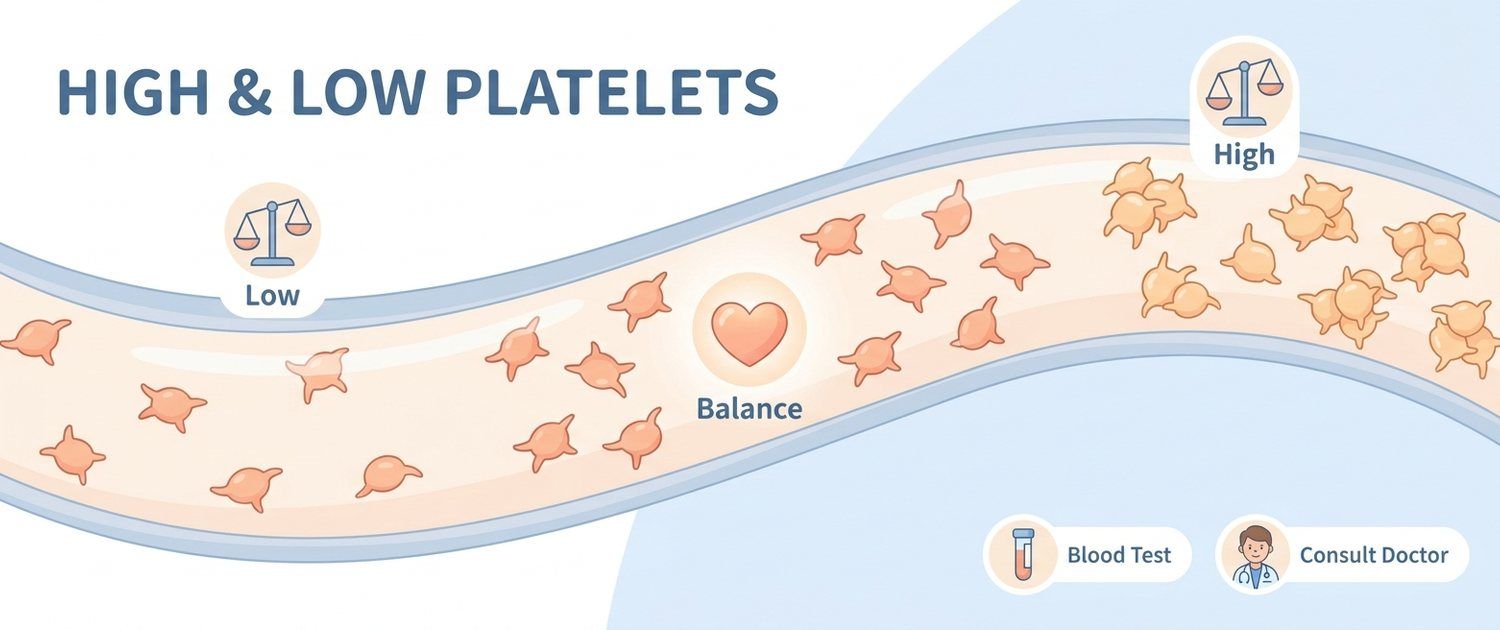
The platelet line on a report frightens people more than most: the word is familiar, it is tied to bleeding and clots, and the asterisk beside the number explains nothing. Yet platelets are the value most likely to be something other than it seems — a share of abnormal results never existed in the body at all and lives only inside the tube. Reading a full report is easier with the general guide: Blood Test Guide.
Platelets are not cells in the full sense but anuclear fragments of giant marrow cells called megakaryocytes. They live 7–10 days, and a third of the total pool sits in the spleen at any moment. That gives three sources of an abnormal platelet count: production in the marrow, destruction in the periphery, and redistribution between blood and spleen.
Below: why iron deficiency raises platelets more often than anything else, how pseudothrombocytopenia differs from the real thing, what separate ranges apply in children and in pregnancy, and when low counts really matter.
Normal platelet count in adults
The reference range is the same for men and women and barely changes with age.
| Value, × 10⁹/L | Name | What it usually means |
|---|---|---|
| 150–400 | Normal | No abnormality |
| 400–600 | Mild thrombocytosis | Almost always reactive: inflammation, iron deficiency, recent infection |
| 600–1000 | Moderate thrombocytosis | Usually reactive, but the cause must be found |
| Above 1000 | Marked thrombocytosis | Bone marrow disease must be excluded |
| 100–150 | Mild thrombocytopenia | Often a normal variant or transient |
| 50–100 | Moderate thrombocytopenia | Needs a cause; bleeding is unusual |
| 20–50 | Severe thrombocytopenia | Bruising and bleeding after injury |
| Below 20 | Critical thrombocytopenia | Risk of spontaneous bleeding |
One caveat about the boundary: some healthy people sit at 130–145 × 10⁹/L for life. That is a familial trait, recognised by one feature — it never changes across years of testing.
Platelet count in children and in pregnancy
In newborns the range is wider than in adults; by one month values climb and stay above the adult range through early childhood.
| Age | Normal, × 10⁹/L |
|---|---|
| Newborn | 150–450 |
| 1 month — 1 year | 180–500 |
| 1–6 years | 180–450 |
| 6–12 years | 160–450 |
| Over 12 years | 150–400 |
So a value of 480 is normal in a one-year-old and a reason to look for causes in an adult. In small children thrombocytosis is almost always reactive: any infection lifts the platelet count for 2–4 weeks. Paediatric reports are covered in Child Blood Test Norms and CBC with Differential.
Pregnancy runs the other way: plasma volume grows faster than cell mass and platelets are diluted. By the third trimester 5–10% of women fall to 100–150 × 10⁹/L — gestational thrombocytopenia, which is safe and resolves after delivery. What does raise concern is a drop below 100 together with high blood pressure, protein in the urine and rising liver enzymes. These physiological shifts are covered in CBC in Pregnancy.
High platelet count: reactive thrombocytosis
Reactive (secondary) thrombocytosis is a normal marrow responding to an outside stimulus, and it accounts for 80–90% of all raised results. The mechanism is shared: inflammation and iron shortage drive interleukin-6 and thrombopoietin, which spur the megakaryocytes.
| Cause | How it is recognised |
|---|---|
| Iron deficiency | Low ferritin, raised TIBC, microcytosis on the indices |
| Acute infection and inflammation | Rising CRP and ESR, a recent illness |
| Chronic inflammation | Rheumatic disease, inflammatory bowel disease |
| Blood loss | Recent surgery, delivery, heavy periods, bleeding |
| Removal of the spleen | Surgery in the history; the rise is permanent |
| Marrow recovery | Rebound after chemotherapy or after stopping alcohol |
| Malignancy | Thrombocytosis as a paraneoplastic sign |
| Exercise, stress | Brief, gone on a resting repeat |
Iron deficiency comes first and is checked first. The logic is plain: it is the commonest cause, the cheapest to test and the most reversible. Look at Ferritin (Iron Stores), Serum Iron Test and TIBC Blood Test — the last rises as stores empty, because the carrier protein is left unloaded. Restoring iron normalises the platelet count within 4–8 weeks with no separate action on clotting.
An important detail: reactive thrombocytosis, even a marked one, does not itself raise the risk of thrombosis. The platelets are functionally normal, there are simply many of them. That separates it from clonal disease, where behaviour rather than number changes.
High platelet count: clonal thrombocytosis
Clonal thrombocytosis is a disease of the marrow itself: megakaryocytes produce platelets independently of need. The main entity is essential thrombocythaemia; the same mechanism appears in polycythaemia vera and primary myelofibrosis, discussed in High and Low Hematocrit.
Underneath is an acquired mutation in a progenitor cell: JAK2 V617F in roughly 55–60% of patients, CALR in 20–25%, MPL in 3–5%. These mutations are not inherited and are not passed to children. Marrow tumours of uncertain behaviour found on workup may be coded as D48 — neoplasm code.
What marks clonal disease out:
- Persistence. The value holds for months and does not move once infection is cleared and iron restored.
- No visible cause. Ferritin is normal, there is no inflammation, there was no blood loss.
- Thrombosis and paradoxical bleeding. At very high counts the platelets consume von Willebrand factor, so excess clotting coexists with bleeding.
- Associated findings. An enlarged spleen, shifts in other lineages, altered red cell indices — read through MCV, MCH, MCHC and RDW.
| Feature | Reactive | Clonal |
|---|---|---|
| Share of cases | 80–90% | 10–20% |
| Cause visible | Yes, nearly always | No |
| Course | Gone within weeks | Holds for months and years |
| Spleen | Usually not enlarged | Often enlarged |
| Thrombosis risk | Not raised | Raised |
| JAK2, CALR, MPL mutations | Absent | Present in 85–90% |
A haematologist makes this diagnosis, not the analyser: it takes a repeat count, ferritin, inflammatory markers, genetic testing and sometimes a marrow biopsy.
Low platelet count: the false one in the tube
Before real causes are considered, the cause that does not exist in the body is ruled out. In 0.1–2% of people platelets clump in the presence of EDTA, the preservative in the standard CBC tube. The analyser sees a clump as one large particle and ignores the rest, so the report shows 40–70 × 10⁹/L in someone whose true platelet count is 250.
This is EDTA-dependent pseudothrombocytopenia. It is recognised by:
- No bruising, no petechiae and no bleeding despite a frightening number.
- An analyser flag for aggregation or clumped platelets.
- Normal values on earlier tests and no change in how the person feels.
The check is simple and belongs first: repeat the draw in a sodium citrate tube (light blue cap), or ask the laboratory to look at a blood film, where the clumps are visible. If the citrate result is normal, there is no disease and nothing further to chase. Skipping this step regularly leads to needless workups and treatment where nothing needs treating.
Causes of low platelets that are real
Once the false result is excluded, the causes fall into three mechanisms.
| Mechanism | Causes |
|---|---|
| Peripheral destruction | Immune thrombocytopenia, drug-induced, infections, antiphospholipid syndrome |
| Reduced production | Marrow disease, B12 and folate deficiency, alcohol, chemotherapy |
| Redistribution to the spleen | Hypersplenism in liver disease and portal hypertension |
What sits behind those lines:
- Viral infection. The commonest reversible cause: EBV, cytomegalovirus, hepatitis, HIV, parvovirus B19, dengue. Platelets fall for one to three weeks and recover on their own. The white differential usually shifts too — see High and Low Neutrophils.
- Drugs. A long list: heparin, some antibiotics, anticonvulsants, anti-inflammatories, quinine. Drug-induced thrombocytopenia typically starts 5–10 days after the drug is begun and resolves after it stops. Stopping a prescription is a decision for the doctor, not the patient.
- Immune thrombocytopenia. Antibodies form against the person's own platelets. In children it usually follows a viral illness and resolves; in adults it tends to be chronic. It is a diagnosis of exclusion — no specific test exists.
- Liver disease. A double hit: the liver makes less thrombopoietin while an enlarged spleen holds more platelets. In Liver Cirrhosis a low platelet count is often the first laboratory sign, long before jaundice.
- B12 and folate deficiency. Usually hits every lineage at once, so platelets fall alongside red cells of strikingly large mean volume.
- Alcohol. Directly suppresses megakaryocytes; values recover over 5–10 days of abstinence.
What to do about a low platelet count
Action depends on depth and symptoms, not on the fact of a low value alone.
- 100–150 × 10⁹/L. No bleeding occurs. Repeat in 2–4 weeks and confirm the value is stable.
- 50–100 × 10⁹/L. No spontaneous bleeding, but a doctor should establish the cause. Ordinary daily activity is generally unrestricted.
- Below 50 × 10⁹/L. Bruises appear after trivial contact. This needs a face-to-face appointment within days, not weeks — and immediately if any of the red flags below is present.
- Below 20 × 10⁹/L. Risk of spontaneous bleeding, including internal. This is urgent.
Red flags — seek care immediately:
- a platelet count below 20 × 10⁹/L on a fresh sample;
- bruises appearing without any injury;
- petechiae — pinpoint red spots on the shins and feet that do not blanch under pressure;
- gums bleeding during ordinary tooth brushing;
- nosebleeds that are hard to stop;
- blood in the urine or black tarry stools;
- in women, periods that have suddenly become much heavier;
- severe headache or confusion with a low count.
What not to do: try to raise platelets with diet or supplements. Food has no meaningful effect on the number, and the lost time works against the person. With a markedly low value the only sensible step is a doctor.
MPV and the clotting neighbours
Next to the number the report prints MPV, the mean platelet volume (roughly 7.5–11.5 fL). It reads as an indirect marker of population age: young platelets are larger than old ones.
- A high MPV with a low count means the marrow is working and releasing large young cells — the picture of peripheral destruction.
- A low MPV with a low count says the opposite: production is suppressed and the marrow is not responding.
- An isolated MPV shift with a normal count carries no diagnostic weight.
The platelet count alone does not describe clotting. The plasma side is covered by other values: Fibrinogen Test as the substrate of a clot and D-Dimer Test as the trace of a clot already broken down. Where vein thrombosis is suspected — coded as I80 — phlebitis and thrombophlebitis — the decision rests on the clinical picture and imaging, not on the platelet count.
Which tests follow an abnormal platelet count
The first step is always the same: repeat the complete blood count, in a citrate tube if the value was low. A single abnormal result without symptoms is not a diagnosis.
| Situation | What else is checked |
|---|---|
| High platelet count | Ferritin, iron, TIBC, CRP, ESR, search for an inflammatory focus |
| Persistent thrombocytosis without cause | Haematology referral, JAK2 and other mutations |
| Moderately low count | Liver panel, viral markers, B12 and folate, review of drugs |
| Markedly low count | Urgent haematology, blood film, marrow if needed |
| Planned surgery | A coagulation panel alongside the platelet count |
If the report is already in hand and the goal is to understand every line together, that is what a lab report review does — it reads platelets in the context of the whole haemogram rather than as a lone figure.
This article is informational. Diagnosis and treatment decisions belong to a doctor.
For informational purposes only
This article is for informational purposes only and does not constitute medical advice, diagnosis, or treatment. Please consult a healthcare professional for medical guidance.