Blood Tests for Weight Loss: Hormones and Metabolic Workup
Reviewed by the LabReadAI medical team
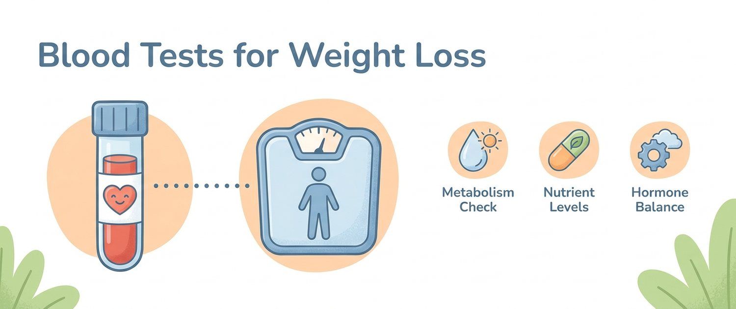
If weight won't shift despite a calorie deficit and exercise, start with eight tests: fasting glucose and insulin with a HOMA-IR calculation, glycated haemoglobin (HbA1c), TSH and free T4, ferritin, cortisol, vitamin D, and a lipid panel. Women with an irregular cycle add sex hormones to screen for PCOS. This is the standard laboratory minimum a doctor uses to assess what specifically is impeding weight loss.
An immediate note of honesty: no test burns fat or removes the need for a calorie deficit. Hormones are not a "weight-loss button". The point of a workup is different — to find a condition that slows metabolism, drains the energy needed to move, or drives fat accumulation: hypothyroidism, insulin resistance, iron deficiency, chronic stress. If such a condition exists and goes unseen, any diet works against a headwind. Tests reveal that wind so a doctor can choose tactics — not so you can treat yourself.
Carbohydrate Metabolism: Insulin, Glucose and HOMA-IR — the Key Link for Weight
This is the core of the workup. In most people with abdominal obesity, weight stalls because of disturbed carbohydrate metabolism — not the thyroid, as is commonly assumed.
Fasting glucose — the baseline screen. Normal is up to 5.5 mmol/L; 5.6–6.9 mmol/L is prediabetes; ≥ 7.0 mmol/L (confirmed twice) is diabetes. But glucose stays normal for a long time even after metabolism is already broken — because the pancreas compensates by flooding the blood with insulin. So glucose alone is not enough.
Fasting insulin — this reveals the hidden problem earlier than glucose. High insulin with normal sugar means cells have stopped responding to it normally and the pancreas is overworking to keep sugar in range. And insulin is the storage hormone: while it is high in the blood, the body is physiologically set to store fat rather than break it down. That is precisely why a person with hyperinsulinaemia loses weight more slowly at the same calorie deficit.
HOMA-IR — a calculated index of insulin resistance from the two values above: HOMA-IR = (fasting glucose × fasting insulin) / 22.5. A value above 2.5–3.0 is usually read as insulin resistance (there is no single cutoff — it depends on population and laboratory). This is the most informative "weight" marker: it explains why weight won't move while glucose is still normal. A full breakdown is in the insulin resistance article.
Glycated haemoglobin (HbA1c) — mean glucose over 2–3 months; no fasting needed and resistant to a single overeating episode the night before. In weight problems it screens out prediabetes (5.7–6.4%) and diabetes (≥ 6.5%), which often accompany obesity and make weight loss harder.
What an abnormal result suggests: high insulin and HOMA-IR with normal glucose — insulin resistance, the most common metabolic brake on weight; high glucose and HbA1c — prediabetes or diabetes already, needing separate tactics.
Thyroid: TSH and T4 — When Metabolism Is Slowed
The thyroid sets the pace of metabolism. When it underperforms (hypothyroidism), the body spends less energy at rest, fluid accumulates, activity drops — and weight rises even without overeating. This is the second most common condition sought when someone complains about weight.
TSH (thyroid-stimulating hormone) — the primary screening test. It is a pituitary hormone that commands the thyroid: when thyroid hormones run short, TSH rises to try to spur it on. Normal is roughly 0.4–4.0 mIU/L. Elevated TSH signals hypothyroidism (see hypothyroidism); low TSH signals hyperthyroidism, which conversely can cause weight loss.
Free T4 — the active thyroid hormone in the blood. It is read together with TSH to separate overt hypothyroidism (TSH high, T4 low) from subclinical (TSH raised, T4 still normal). It is convenient to order them together as a thyroid panel.
A dose of realism is needed here. Real hypothyroidism does slow metabolism, but the "weight–thyroid" link is overrated: most people with obesity have a normal TSH, and obesity itself can modestly raise TSH — meaning sometimes the thyroid is not to blame for the weight, but the excess weight is nudging TSH. A systematic review and meta-analysis (Frontiers in Immunology, 2019) showed obesity raises the risk of hypothyroidism 1.86-fold and of overt hypothyroidism 3.21-fold, largely through adipose-tissue inflammation rather than the reverse. So treating hypothyroidism normalises metabolism but on its own rarely produces large weight loss — observational data on levothyroxine therapy confirm this.
What an abnormal result suggests: high TSH (especially with low T4) — hypothyroidism as one cause of slowed metabolism; normal TSH — the thyroid can be crossed off the list of weight "culprits".
Iron and Energy: Ferritin — Why There's No Strength to Move
Weight loss rests not only on diet but on movement — and movement needs energy. Hidden iron deficiency quietly steals exactly that: chronically tired, breathless on the stairs, unable to muster energy for a workout — and the person blames laziness or age, though the cause is in the blood.
Ferritin — the iron-storage protein; it shows the body's iron reserves before haemoglobin falls. It is the most sensitive marker of hidden deficiency. Values below 30 µg/L indicate depleted stores even with a formally normal complete blood count. Low ferritin is especially common in women of reproductive age due to menstrual losses. Thresholds are covered in the ferritin article, and full iron-metabolism assessment comes from the iron panel.
An important caveat: ferritin is also an inflammatory protein, so in obesity, infection or inflammation it can be falsely elevated and mask a deficiency. It must be read in context, not from a single number.
What an abnormal result suggests: low ferritin — depleted iron stores, a likely cause of fatigue and lack of energy for activity; high ferritin alongside obesity — more often an inflammation marker than iron excess.
Cortisol and Vitamin D: Stress, Abdominal Fat and Metabolism
Cortisol — the stress hormone. In chronic stress its excess drives fat specifically to the belly (abdominal, "central" obesity), intensifies sugar cravings and raises blood sugar. Baseline screening is morning blood cortisol (peak secretion), or evening salivary / 24-hour urinary cortisol to assess the rhythm. Sharply and persistently elevated cortisol with a characteristic "moon" face, stretch marks and muscle weakness is a reason to rule out the rare but serious Cushing's syndrome (hypercortisolism); the European guideline on endocrine workup in obesity (Eur J Endocrinol, 2020) names it explicitly as a reason to refer to an endocrinologist.
Vitamin D (25-OH vitamin D) — its deficiency is very common in obesity: adipose tissue "sequesters" the vitamin, lowering its availability. Vitamin D deficiency is not a direct cause of excess weight, but it is associated with insulin resistance, muscle weakness and low mood — backdrops that make an active lifestyle harder to build. Deficiency is a level below 20 ng/mL (50 nmol/L).
What an abnormal result suggests: high cortisol — a stress mechanism of belly-fat gain, and in rare cases hypercortisolism; low vitamin D — a common deficiency worth correcting as a backdrop, but not as a "cause" of weight.
Lipids and Liver: Metabolic Syndrome and Fatty Liver
These tests are not about weight itself but about its metabolic consequences — how far the extra kilograms have already hit the vessels and liver.
Lipid panel — total cholesterol, LDL, HDL and triglycerides. The characteristic "weight" pattern is high triglycerides and low HDL: this is the atherogenic dyslipidaemia that accompanies insulin resistance and is one of the criteria for metabolic syndrome.
Liver enzymes (ALT, AST) — in abdominal obesity ALT is often elevated: an indirect sign of non-alcoholic fatty liver disease (NAFLD), where excess insulin drives fat into liver cells. NAFLD is present in most people with marked metabolic syndrome and itself sustains insulin resistance, closing the loop.
What an abnormal result suggests: high triglycerides and low HDL — metabolic syndrome and insulin resistance; elevated ALT — likely fatty liver, another brake on metabolism.
Sex Hormones in Women: PCOS
In women with excess weight, an irregular cycle, acne and excess male-pattern hair growth, a frequent cause is polycystic ovary syndrome (PCOS). It is rooted in the same insulin resistance that both hinders weight loss and raises male hormones. The baseline panel includes testosterone (total and free), SHBG, LH and FSH, plus prolactin to exclude other causes of cycle disruption. These hormones must be read against the cycle day and always with a doctor — interpretation here is non-trivial.
What an abnormal result suggests: raised male hormones and a characteristic LH/FSH profile in a woman with an irregular cycle — likely PCOS, in which working on insulin resistance specifically often kick-starts weight loss.
Summary Table: Marker → What It Assesses → What an Abnormal Result Suggests
| Marker | What it assesses | What an abnormal result suggests |
|---|---|---|
| Fasting glucose | Current sugar level | High — prediabetes/diabetes |
| Fasting insulin | Load on the pancreas | High with normal sugar — hidden insulin resistance |
| HOMA-IR (calculated) | Insulin resistance | > 2.5–3.0 — body set to store fat |
| HbA1c | Sugar over 2–3 months | ≥ 5.7% — prediabetes; ≥ 6.5% — diabetes |
| TSH | Thyroid function | High — hypothyroidism, slowed metabolism |
| Free T4 | Active thyroid hormone | Low with high TSH — overt hypothyroidism |
| Ferritin | Iron stores | Low — fatigue, no energy to move |
| Cortisol | Stress level | High — belly fat, sugar cravings |
| Vitamin D (25-OH) | Vitamin D status | Low — backdrop for IR and muscle weakness |
| Lipid panel | Cholesterol and triglycerides | TG↑ + HDL↓ — metabolic syndrome |
| ALT / AST | Liver status | ALT↑ — likely fatty liver |
How to Take These Tests Correctly
- Strictly fasting (8–12 hours without food, water allowed) — glucose, insulin, lipids. Even a small snack distorts the result and nullifies HOMA-IR.
- In the morning — cortisol (peak secretion) and, where possible, the whole hormone block.
- HbA1c and TSH need no fasting — they can be taken at any time.
- Sex hormones in women — timed to the cycle day (usually days 2–5), as prescribed by a doctor.
- Do not test hormones during acute illness, severe sleep deprivation or a crash diet the day before — the result will be distorted.
What to Do with the Results
The collected tests are a map, not a diagnosis — and certainly not a prescription. The same "high insulin" in a young woman with PCOS and in a man with long-standing obesity calls for different tactics, and reference ranges depend on the laboratory, sex, age and clinical picture. So leave the interpretation and — above all — the decision on what to do next to an endocrinologist or GP: prescriptions, dosages and medications are outside the scope of this article and outside the realm of self-treatment.
LabReadAI helps with the first step — understanding your numbers before the doctor's visit: upload your test report and the analysis will explain what each marker means in your context and what to flag for your doctor. This makes the conversation in the consulting room faster and more focused, but does not replace it.
Further reading: how insulin resistance hinders weight loss and what a doctor does about it — in the insulin resistance article; why obesity itself sustains hormonal shifts — there too; and how individual disturbances combine into metabolic syndrome — in the dedicated review.
This article is for informational purposes only. Interpretation of tests, diagnosis and treatment are the responsibility of an endocrinologist or general practitioner.
For informational purposes only
This article is for informational purposes only and does not constitute medical advice, diagnosis, or treatment. Please consult a healthcare professional for medical guidance.