What Does a Blood Test Show: Guide to Every Key Result
Reviewed by the LabReadAI medical team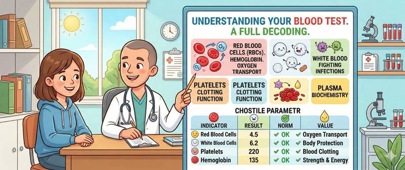
A sheet of numbers, upward and downward arrows, and cryptic abbreviations — that is what a blood test result looks like to most patients. What does it actually tell a doctor, and how are those figures connected to health and disease? This article is a comprehensive breakdown of all the main categories of blood tests, from a routine full blood count to tumour markers. Before taking any of them, correct preparation matters — detailed rules are covered in the blood test preparation guide.
Complete Blood Count: Cells, Haemoglobin and Immune Signals
The complete blood count (CBC) is the most widely ordered investigation in medicine. It evaluates three cellular lines and provides an initial snapshot of the whole body's condition.
Red cells and haemoglobin. Haemoglobin is the oxygen-carrying protein inside red cells. Its fall — anaemia — presents as fatigue, pallor, and breathlessness. Normal range: 130–170 g/L in men, 120–160 g/L in women. MCV — mean red cell volume — is the critical differentiator: small cells point to iron deficiency; large cells to vitamin B12 or folate deficiency. This is the first step in the differential diagnosis of iron deficiency anaemia and other forms.
White blood cells. Leukocytes are the cells of immune defence. An elevated count signals infection, inflammation, stress, or leukaemia; a low count indicates bone marrow suppression, severe viral illness, or a drug side effect. Normal range: 4–9 × 10⁹/L. The differential count (extended CBC) clarifies which cells are affected: neutrophils in bacterial infection, lymphocytes in viral illness, eosinophils in allergy and parasitic disease.
Platelets. Platelets carry out primary haemostasis — the first seal on a damaged vessel. A count below 100 × 10⁹/L raises a bleeding risk; a fall below 20–30 × 10⁹/L carries the threat of spontaneous haemorrhage. Elevated platelets often accompany inflammatory conditions or iron deficiency.
A single CBC does not establish a diagnosis — it points in a direction. Low haemoglobin, small red cells, and a high platelet count together almost certainly mean iron deficiency; simultaneously low counts across all three lines (anaemia, leucopenia, thrombocytopenia) is a signal to investigate the bone marrow.
Biochemistry Panel: Liver, Kidneys and Metabolism
Biochemistry is the principal tool for assessing organ function. It is ordered in virtually every chronic disease workup.
Liver function tests. The liver function test panel measures several markers with distinct clinical meanings. ALT and AST are enzymes that "leak" from damaged hepatocytes into the blood — their elevation marks active liver cell destruction in hepatitis, fatty liver disease, or drug-induced injury. GGT is particularly sensitive to alcohol and cholestasis; bilirubin reflects haemoglobin processing and bile flow; albumin measures the liver's synthetic capacity — its ability to produce proteins. Albumin below 30 g/L indicates severe hepatic failure or major systemic inflammation.
Kidney function markers. The kidney function test includes creatinine and urea. Creatinine is a waste product of muscle metabolism cleared by the kidneys: its accumulation indicates falling glomerular filtration rate (GFR). GFR — calculated from creatinine — is the primary staging marker for chronic kidney disease. Urea is less specific and is influenced by diet and tissue breakdown, but in combination with creatinine it helps track renal function over time.
Glucose. Glucose in a biochemistry panel screens for diabetes and prediabetes. Normal fasting value: below 5.6 mmol/L. Values of 5.6–6.9 indicate prediabetes; 7.0 or above on two measurements meets the diagnostic threshold for diabetes. Glucose is interpreted alongside C-peptide — which quantifies the pancreas's residual insulin output — when differentiating type 1 from type 2 diabetes is clinically necessary.
Hormone Tests: Thyroid, Diabetes and Reproductive Health
Hormones regulate metabolism, reproduction, growth, and stress responses. Their abnormalities produce symptoms that frequently mimic other diseases — from fatigue to depression.
Thyroid hormones. The thyroid panel — TSH, free T4, and where indicated free T3 with antibodies — is the essential set for diagnosing thyroid dysfunction. TSH is the most sensitive marker: its elevation means the pituitary is signalling the thyroid to work harder — a sign of hypothyroidism. A suppressed TSH indicates a hyperactive gland. Free T4 clarifies whether hormone levels are genuinely low (primary hypothyroidism) or the problem lies in the pituitary (secondary). For thyroid nodules, calcitonin is always added — the marker of medullary thyroid cancer.
Sex hormones and adrenal markers. The hormone panel covers FSH, LH, oestradiol, testosterone, progesterone, and prolactin. It is ordered for menstrual irregularities, infertility, menopausal symptoms, and hypogonadism. Every hormone has a strict dependency on the time of day and day of the cycle — results can only be interpreted with these parameters accounted for.
Some conditions require simultaneous assessment of multiple systems: polycystic ovary syndrome combines an FSH/LH imbalance with insulin resistance, so sex hormones and fasting insulin are measured together.
Lipid Panel: Cholesterol and Cardiovascular Risk
Cardiovascular disease is the leading cause of death worldwide, and its primary laboratory predictor is a disturbed lipid profile. The lipid panel provides four key measurements.
Total cholesterol is the sum of all lipoprotein fractions — meaningful only in context. "Bad" LDL deposits in arterial walls and is the main treatment target: its goal level ranges from < 3.0 to < 1.4 mmol/L depending on individual cardiovascular risk. "Good" HDL carries cholesterol back to the liver — a protective factor; its fall below 1.0 mmol/L in men is independently hazardous. Triglycerides are neutral blood fats; a rise above 5.6 mmol/L sharply elevates the risk of acute pancreatitis.
A disturbed lipid profile is the primary laboratory marker of atherosclerosis. This is why a lipid panel is recommended as a screening test for all adults over 40, and tested every 3–6 months in patients on statin therapy to monitor treatment effectiveness.
Inflammation, Autoimmunity and Thrombosis Markers
This group of tests is critical for diagnosing systemic processes and thrombotic complications.
Inflammatory markers. C-reactive protein is a fast and accurate marker of active inflammation — responding within 6–12 hours. ESR is a slower, indirect marker that normalises weeks after recovery. Both rise in infections, autoimmune diseases, malignancies, and after trauma. A persistently elevated ESR without an established cause warrants targeted investigation — covered in detail in the elevated ESR article. When rheumatoid arthritis is suspected, rheumatoid factor and anti-CCP antibodies are added to CRP and ESR.
Coagulation and thrombosis markers. The coagulation panel assesses the overall haemostatic system. Fibrinogen is simultaneously an acute-phase protein and the raw material for clot formation: its sustained elevation is an independent cardiovascular risk factor. D-dimer is a product of fibrin breakdown — its rise indicates active clot formation somewhere in the circulation. A normal D-dimer at low pre-test probability virtually excludes pulmonary embolism.
Cardiac markers. Troponin is the primary marker of cardiomyocyte death: its rise within 1–3 hours of chest pain onset is the criterion for acute myocardial infarction. A normal high-sensitivity troponin at 3 hours from symptom onset has a high negative predictive value for ruling out infarction.
Tumour Markers, Vitamins and Electrolytes
Tumour markers. The tumour marker panel is not a population screening tool: most tumour markers rise in benign conditions and lack sufficient specificity for independent diagnosis. The exception is calcitonin: its elevation above 100 pg/mL in the setting of thyroid nodules is highly specific for medullary thyroid cancer and requires immediate oncological evaluation. Tumour markers are primarily used for post-treatment surveillance, not initial cancer detection.
Vitamins and micronutrients. The vitamin panel covers both fat-soluble and water-soluble vitamins. Vitamin D deficiency affects 50–80% of people in northern latitudes and is associated with immune impairment, osteoporosis, and muscle weakness; the optimal level is above 75 nmol/L. Vitamin B12 is essential for the nervous system and red cell production: its deficiency in vegetarians and older adults often presents as depression or early cognitive decline. Ferritin — the body's iron storage protein — falls long before haemoglobin drops and is the first marker of latent iron deficiency. The full iron panel is ordered when a more complete assessment of iron status is needed.
Electrolytes. The electrolyte panel measures sodium, potassium, chloride, and bicarbonate. Calcium governs neuromuscular transmission and bone density: its fall causes cramps and tetany; its rise causes weakness and cardiac arrhythmias. Sodium determines the body's water balance: a critical fall below 125 mmol/L can lead to cerebral oedema. Potassium controls cardiac rhythm: values above 6.5 or below 2.5 mmol/L threaten life-threatening arrhythmias and require emergency correction.
When Blood Test Results Need Urgent Medical Attention
Most abnormal results warrant a planned consultation — not immediate alarm. But certain values require emergency action.
Call emergency services immediately if: glucose exceeds 16 mmol/L or falls below 2.5 mmol/L on a fasting sample — possible hypoglycaemic shock or diabetic ketoacidosis; in patients with type 2 diabetes these thresholds should be stored in memory; troponin is elevated with chest pain — a sign of acute infarction; potassium falls below 2.5 or exceeds 6.5 mmol/L — imminent cardiac arrest risk; haemoglobin falls below 70 g/L — severe anaemia often requiring hospitalisation; fibrinogen drops below 1.0 g/L in the setting of active bleeding — DIC syndrome; calcitonin exceeds 100 pg/mL with a thyroid nodule — urgent oncology referral.
A planned consultation is indicated when: any marker is consistently outside the reference range on two or more successive tests; a result has changed substantially from a previous value despite correct preparation; a serological test (rheumatoid factor, anti-CCP antibodies, tumour markers) returns strongly positive — a single test never establishes a diagnosis; it requires clinical interpretation.
Blood tests are a physician's tool. Understanding their language is useful. Interpreting them without clinical context is dangerous. The same number in two different patients can mean two entirely different things.
This article is for informational purposes only and does not replace medical consultation. Result interpretation and diagnosis are made by a qualified physician.
For informational purposes only
This article is for informational purposes only and does not constitute medical advice, diagnosis, or treatment. Please consult a healthcare professional for medical guidance.